Oncogenes
are important for the classification of cutaneous lymphomas,
leukaemia’s and
other cancers. The pathologist Peyton Rous observed 1911 in ill
chickens the
transfer of cancer over the injection of tumour infected fluidics into
the body
of healthy chickens [1]. The oncogene src leads for healthy chickens to
cancer
(tumour sarcoma). This oncogene src is also infectious or contagious
for human cancer
that has been proven sufficiently. The HIV infection - over the blood
is a mechanism
of gene transfer from an ill body to a healthy body via oncogens (DNA
or RNA).
The src - oncogene is able, to produce an unlimited cell growth. The
transfer
of oncogens is not the same as a viral infection. HIV
is not a virus but the gene transfer via blood of an e.g. myc
oncogene that initialize at first a pre stage of cancer as the cause
of
HIV and HIV- PCR-, ELISA-test results.
The pathologist
Peyton Rous [1] observed
1911 in chickens the transfer of cancer by body fluidics. The injection
of the
extract with chicken sarcoma led to the illness of healthy chicken.
Healthy
chicken developed within a view months or years a cancer of sarcoma.
Rous thought
that was a virus (RSV), which is the
cause for this cancer. Rous 1966 received the Nobel Prize medicine for
his
discovery. Only a gene or oncogene v-src
or RSV con trigger the cancer and
requires no virus for the transfer. The DNA
of the src yields of a restriction
enzyme
of the DNA from the bacteria phage.
The replication of a v-src DNA is cloned by a bacteria phage. The DNA can cut
in pieces and recombined with plasmids, which are increased in
bacteria cells. Different DNA are
cloned by the bacteria with a certain fragment of the Phage- DNA and carries also the DNA
of the RSV (src)
oncogene in
it. The transfer of these RSV gene
fragments in the DNA by cells
transforms lead to a cell degeneration and tumour grow. A weight has
marked.
The oncogene src has a weight of 60kD or called p60-src
The proto-oncogenes (c-src)
is also in healthy cells, that converts to virale oncogenes (v-src) the origin for the tumour. Many
oncogenes
have been discovered:
Proto oncogene und cancer by
animals
c-abl Abelson- mouse
leukaemia
c-bcl B-cell lymphoma
c-erb Erythroblasts
c-fos
FBJ- mouse-osteo
sarcoma
c-fps Fujinami-PRCII-avian
sarcoma
c-int insertions activate onc of
mouse mamma
carcinom
c-jun avian
sarcoma-Virus 17-Gen (junana)
c-met methyl-nitroso-guanidine
treated human
osteo sarcoma
c-mil avian
gel-Mill-Hill-2-Retrovirus
c-mos Molony-
Mouse
sarcoma
c-myk Myelozytomatose
c-myb
avian-myeloblastose
c-ras Kirsten-Ratt
sarcoma
c-rel avian-retikuloendotheliose
c-src Rous-sarcoma
c-sis simian
sarcoma
Proto oncogene of human cancers
c-abl leukaemia
c-bcr B-cell
lymphoma
c-my Burkitt lymphoma
c-bcl follicular
B-cell lymphoma
Cancer associated with oncogene
Virus Genome
cancer
RSV
v-src
sarcoma
ALV
B-cell- lymphoma
MC 29
v-myc
myeloid leukaemia
MLV
T-cell-lymphoma
Abelson
v-abl
B-cell- lymphoma
HIV can better understand to know
the
history for the most important medician R.C. Gallo [6]. He has at first
analysed leukaemias because his sister was died on this illness. He
found the HLTV-I virus [6] that he
has held for a
virus with the cause of leukaemia although HLTV-I
is found in the blood of 5% in all leukaemias. Cutaneous B-Cell and T-
cell-
lymphomas have also been investigated by Gallo [7]. The
Kaposi sarcoma is a cutaneous B-cell lymphoma with a direct connection
of HIV and AIDS.
Today, Kaposi sarcoma and other tumours are the second stage
of HIV and AIDS.
HIV corresponds directly to the HLTV- III virus and is derived from the
leukaemia virus HLTV-I.
AIDS antiviral medicaments such as
Zitrovir AZT (originally a
cytotoxic) decrease the number of leukocyte and diminish the powers of
resistance of the body. The only once definition of AIDS
is the reduction of the relationship CD4/CD8
between the T-helper- to T- cytotoxic cells CD4/CD8.
The decreasing of leukocyte CD4 or
T -helper cells has the cause in
the antiviral therapy and the following immunodeficiency. The transfer
of
oncogenes into another
body can trigger
a tumour that repeatedly was proved. In this sense is HIV
the result of exchanged fluidics, particularly blood of two
bodies. After an infection with an oncogene it appears often a
cutaneous tumour
such as Kaposi sarcoma in the second stage of AIDS
in proximally 30-40% of all HIV–positive
cases. The important knowledge of HIV
is, that cancer can be initiated by an infection over the
exchange of body fluidics such as blood.
Current experiences show, that
no HIV - virus can be selected by
Virchows
law. The original methods after R.C. Gallo do not find a virus with
scientific methods
of the virology. If HIV is
considered
as a pre stage of cancer we can cancel the virus theory. The
argumentation for HIV would be
better for the clarification
and is not as dangerous as a deathly illness. The time after incubation
of a
viral HIV infection is too long.
The
HIV test for AIDS
shows more connectivity to cancer and leukaemia or cutaneous
B-cell-lymphoma NHL and the Kaposi
sarcoma.
.
The main protein
of the HIV- infection is a gene
segment with a
weight of 24000 Dalton (24 kD = p24)
that we can also find in cutaneous B- cell- lymphomas [3]
and cutaneous
T-cell lymphomas- and leukaemias.
The HIV test is a genetic PCR analysis and was investigated by
R.C. Gallo [1].
The solution of this problem
could be the feature
or ability of the v-abl oncogene to
initialize leukaemia or B-cell lymphoma by the v-myc
oncogene. Oncogenes
can
infect a body by an infection over the blood, but it is not an
infection of a
real virus. A HIV-infection is than
a
transfer of an oncogene via the blood into another body and a pre stage
of
cancer. The T-cell-infection of CD4+
is a normal reaction of T-cells after an infection as the immune
response of
cancer but it s not an immunodeficiency.
The essential
difference between a
virus and a DNA- or RNA
oncogene is that an oncogene leads
to cell growth a virus replicates in a cell for a real infection. An
oncogene
can triggers a point mutation for cell growth (increased cell
division). The
diagnosis HIV positively can be
examined as an
initial pre stage of cancer
with the p24, p41, gp120 protein.
.
CD4
and CD8 are markers
of
T-cells lymphomas- and leukaemias
are important in the
later stage of HIV.
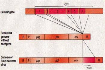
Fig. 1
v-src- and pre- c-src- oncogene [3]
The progress of cancer is often
visible by a
translocation or displacement of cncogenes in the chromosomes.
Leukaemia. is an
example for the translocation of oncogene abl
from chromosome 9 to chromosome 22 and the translocation of oncogene sis from chromosome 22 to chromosome 9.
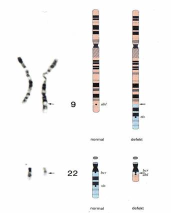
Fig. 2 chromosome
–translocation
by the oncogene abl und sis [3]
The conclusion of the previous
statements about
HIV is, that is more a cancer or the initial pre stage of cancer, than
a virus
infection. The skin of HIV-patients
shows in the later stage of HIV a Kaposi sarcoma.
An example for HIV and
Kaposi sarcoma are
Now, we can
understand that
oncogenes can be transferred by exchanging blood of infected patients.
This
idea is surprising, we
must trust
ourselves with an infection or transfection of oncogenes for HIV and
cancer
A real picture of
a HIV virus does
not exist, because picture in Fig. 3 shows cells of blood. An oncogene
has the feature
of gene transfer via the blood from the infected- to the heath body.
The transfer
of an oncogene initializes the pre stage of a tumour that we can see in
the p24- HIV-test. HIV transfer
oncogenes by
exchanging blood or fluidics between two bodies. The theory of a HIV
virus or
Retrovirus (RNA) must take leave,
since a gene transfection is the cause of HIV. Rous take at first the
idea of a
virus by the transfer of oncogene src,
but he did not have an electron microscope in the year 1911 to see a
virus. All pictures
in Fig. 3 show cells of blood with
but no typical virus structure. All pictures are the same as cells for
leukaemia (HLTV-I)., lymphoma (HLTV-I) and HIV
(HLTV-III).
Fig. 3 HLTV I,
HLTV II, HLTV III (HIV) - virus [6]
Modern analytic
used molecular
biologic methods, the PCR- and ELISA test. Genetic investigation makes
it possible to classify a gene via the length or weight in kilo
The reliability of
a genetic test
as PCR or ELISA
is low with a slight percentage of probability. The PCR-
HIV test is looking for a gene
sequence with a weight of 24000
HLTV
I (leukaemia and cutaneous
lymphoma) is the base for the HIV – virus, both have a
connection
with cancer. Oncogenes are exchanges by the blood as c-abl
that can be found in leukaemia, c-bcr
in B-cell lymphoma and c-myc
in Burkett lymphoma and in HIV in
the later stage.
Oncogenes are an
explanation for a HIV infection via
the blood or the translocalization
of oncogenes in chromosomes. HIV is
an
initial stage of cancer. The immunodeficiency of HIV
associated with the ratio CD4/CD8
of lymphocyte which is the result of a therapy of many years with
cytotoxic
antiviral medicines like as AZT or
Zitrovir .
Fig. 4
Western blood test (ELISA-PCR) of HLTV- III
(HIV) antibodies
and molecular
weights in Kilo
B- cutaneous, lymphoma- patient, C-
positive and negative
homo sexual patient [6]
The definition
of HIV - after
R.C. Gallo
The Adult T-cell leukaemia was
indicated as the
HLTV – I virus in
HLTV-I,
and a TCGF protein
(tumour grow factor) was first found in the blood of black patients of
the
Fig.
5 HLTV- I, HLTV
– II and HLTV- III = HIV
Genome with
LTR,
gag, pol, env und pX cloning
with
EColi bacteria
HLTV-III or HIV
is defined by R.C. Gallo as infection with the HIV
- virus and the destruction of T4 -
helper cells. HIV appears
accordingly at the risk group of homosexuals,
consummates of drugs, haemophilia’s, Haitian and children of
the risk groups.
The surface molecule CD4 of the T4- leukocyte is infected, also CD8 of the T8
leukocyte
The HIV
main protein p41 is found in the
Genome of HLTV-III (HIV) of risk
groups. HIV-AIDS patients are tested for the p24-protein.
The ALV
oncogene belongs after Gallo to the HTLV
family. All HLTV-I,
HLTV-II, HLTV-III
(HIV) contain the p24
– protein as in leukaemia, lymphoma
and HIV.
HTLV and HIV
cellines
Cellines [7] are necessary
for genetic investigations of
cells and genetic tests such as PCR
and ELISA that are important for HIV and cancer. HIV
cellines are coming from the blood of a patient [13] with cutaneous T-cell lymphoma.
The celline H9 and HUT 78 are used for HIV
tests.
HTLV-I
has
the TCGF
= T-Cell-growth factor, the ELISA
test with these cellines H9 or HUT 78 indicates p19
and p24 proteins
and the
antigen CD4+
The
celline HTLVCR
was
established from a 28- years old black man with a cutaneous
T-cell lymphoma (mycosis
fungoides).
The
celline HUT 102 was established
from
the lymph nodes- und CTCL-3 and
peripheral blood of the 28- years old black man with cutaneous
T-cell lymphoma (mycosis
fungoides).
The
celline HUT 78 was established from
a patient with a cutaneous T-cell lymphoma
(Sezary lymphoma), the PM1 celline is derived from HUT 78. The ELISA
test shows p24 und
gp120. The T- cells of PM1 shows the CD
marker CD3, CD4+, CD8-,
CD26 und HLA-DR+.
The
celline H9 is cloned from HUT 78 and is used for the isolation
and production of HIV proteins The celline PM1
is also a HUT78
T-cell line CD4+ clone with the
genetic framework HXB2 and gp120 and gp41 [18].
CD4+ is in
infected T-cells of a cutaneous
T-cell lymphoma (HUT78)
HTLV-III
(HIV) has
no
TCGF, the HIV- ELISA- test shows p24, p41
and the antigen CD4+
HIV and
Oncogenes
Between HIV and oncogenes is a
connection for specific
cancers. Such proteins which are important for the cell nuclides were
investigated in [13]. They are in connection with nuclide-protein-
complex.
Based on amino acids sequences by known nuclides or targets, peptide
was tested
with nuclear target potential on it, whether they can trigger an
infection
through the transfer in the cell. The control of an infection can be
ascertained by reporter - genes PK.
The sequences of the c-myb target potential p53
and c-erb-A oncogene build a hybrid
in the cell - nuclides, which can be identified by reporter -genes PK.
The HIV-tat
protein fuses with the target nuclides of the cell. The blood of HIV- infected patients have following
oncogenes: SV-40, tumour suppressor
gene
p53, c-erb-A, c-myb, c-myc, p53, myc-tat.
T-Cell-Receptor TCR and the CD-marker
CD28 are necessary
for
the activation of primary CD4-T-cells
that was investigated. The expression of the c-myc
protein that be initialized by the stimulation of CD4–T-helper-cells
was investigated in
[14]. Cyclosporine hind the
nuclear
import of the HIV-1 -DNA but also
the expression of the c-myc proteins.
It would be tested further whether the oncogene c-myc
is necessary for the nuclide import of HIV-1-
DNA. It would be tested
the function of the
HIV-1 -DNA over the activation of
primary CD4-T-cells
und des T-cell-
receptor TCR und CD
28 and understand
the expression of the c-myc
protein as the stimulation of CD4–T-
cells.
Obviously the c-myc is an essential
part of the total HIV- genome for
an
infection of the HIV- DNA.
1. Targeting
Proteins Class A [13]
p126
K-K-K-R.K-V-E
SV40 large T 2
p279
P-K-K-A-R-E.V
Polynoma
large T 2
A1-P T-K-R-K-G-S
SV40 VP1 26
p3l6 N-K-K-K-R.K-L
SV40 VP2 21
p120 A-A-K-R-V-K-L-D Human c-Myc 5
2. Protein sequences sub
cellular distribution of
PK (Pyruvate Kinase fusions) [13]
c-Erb-A A.
G22-K-R-K-R-K-S
NT 32
B. S127-K-R-V-A-K-R-K-L
Nuclear
and
C. S251-H-W-K-Q-K-R-K-F NT (10-3076)
c-myb
p521
L-L-K-K-1-K-Q Nuclear and
c-myc
p387
-Q-K-K-I-K-S Nuclear (>95%) 34
p53 p416 Q - P - K - K - K - P Nuclear
HIV- tat G46-R-K-K-R-R-Q-R-R-R-A-P
Myc-tat P-A-A-K-R-V-K-L-D-Q-R-R-R-A-P
Abbreviations
ALV
= Avian
Leucosis Virus
RSV
=
Rous
Sarcoma Virus
HIV = Human
Immunodeficiency Virus
HTLV =
Humane T-Cell Leucosis Virus
MLV
=
Mouse Leucosis Virus
PCR
= Polymere
Chain Réaction
PK
=
Ppyruvate
Kinase fusions
kD
= Kilo
Dalton – molecularly
weight
Literature
[1] Rous, P,
Transmission of a
malignant new growth by
means of a cell-free filtrate, J. Am.
Med. Assoc.,
1911, 56,
198-201
[2]
Molekularbiologische und biochemische
Charakterisierung der
reversen Transkriptase
von
Rous Sarcoma Virus,
Dissertation, Universität
Bochum, Max-Planck-Institut für
Molekulare
Physiologie Dortmund
[3] Vamus
H. Weinberg R. A.
Gene
und Krebs
Spektrum 1992
[4]
Passarge E.
Taschenatlas der Genetik
Thieme Stuttgart 1994
[5] Keller
R.
Immunologie Immunpathologie
Thieme Stuttgart 1994
[6]
Gallo R.C. et. al. The Human T-cell Leukemia-Virus
Family, Adult T-cell Leukaemia, and
Hämatol.
Blutransf.
Vol
29 Science 1984;
224(46-48) 500-503
[7] Gallo
R.C. et. al.. Human
T-cell leukaemia-
lymphoma virus (HTLV) is in T but not in
B
lymphocytes from a patient with cutaneous
T-cell
lymphoma, Proc. Natl. Acad Sci
Science, Vol. 79, pp. 5680-5683,
September 1982
[9] Gallo
R.C. et. al. NATURAL
ANTIBODIES TO
THE HUMAN T-
CELL LYMPHOMA VIRUS
WITH CUTANEOUS
T- CELL
LYMPHOMAS J:
Exp. Med. The Rockefeller
University Press Volume
154 August 1981 333-346
[10] Gallo
R.C. et. al. IN
VITRO CELLULAR
TROPISM OF HUMAN B LYMPHOTROPIC
VIRUS HUMAN HERPESVIRUS
J: Exp. Med.
The
1988 1659-1670
[11]
Diepgen, T., Yihune, G. et al
Dermatology
Online Atlas
[12] HIV Molecular Immunology
2006/2007
Theoretical Biology and Biophysics Group
T-10,
Mail Stop K710
Los Alamos National
Laboratory, Los Alamos,
New Mexico
87545 U.S.A.
[13] Gallo
R.C. et. al. Detection and isolation of
type C
retrovirus particles from fresh and
cultured
lymphocytes of a patient with
cutaneous T-cell
lymphoma
[14] Gallo
R.C. et. al. Detection, isolation
and
continuous production of cytopathic
retroviruses
(HTLV-III) from patients with AIDS and
Pre-AIDS
Science, VOL. 224, April 1984
[15]
Gallo R.C. et. al. Serological Analysis of a
Subgroup of Human T-Lymphotropic Retroviruses
(HTLV-III) Associated with AIDS
Science, VOL. 224, April 1984
[16] Fenyö
E.M., Äsjö B. Growth of
the (HTLV-III)
Strain of the Human
Immunodeficiency Virus in
Different Cell Types
Haematology
and Blood Transfusion Vol. 31
[17] Gallo
R.C. et. al. Expression of cellular
homologues of retroviral onc genes in
human
haematopoietic cells
Proc.
Natl. Acad Sci
Vol.79, pp. 2490-2494, April
1982
[18] Gallo
R.C. et. al. Growth of Macrophage-Tropic
and Primary Human Immunodeficiency
Virus
Type 1 (HIV-1) Isolates in a Unique CD4+
T-cell
Clone (PM1): Failure To Downregulate
CD4 and
To Interfere with CD4-Line-Tropic HIV1
Journal of Virology, June 1995, p.
3712-3720
HIV B-Cell-Kaposi
sarcoma
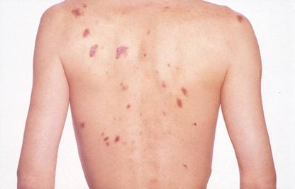
Fig. 6 HIV- B-Cell Kaposi sarcoma
[11]
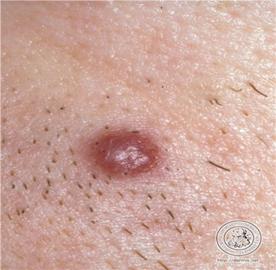
Fig. 7
HIV- B-Cell -Kaposi
sarcoma [111
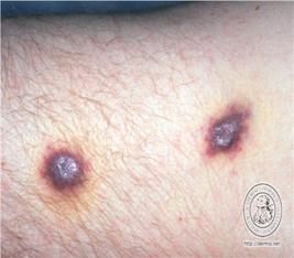
Fig.8 HIV-
B-Cell- Kaposi sarcoma [11]
Fig. 9
HIV Virus Genom
Fig.
10 HIV- Epitope
– mass spectra [12]